
Echocardiogram in Dubai: Types, Procedure, Preparation, Results and Follow-Up
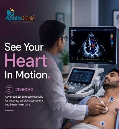
An echocardiogram (echo) is an ultrasound test that creates moving images of the heart. It helps a cardiologist assess the heart’s structure, pumping function, valves and blood flow. At Apollo Clinic Dubai, patients can discuss whether an echocardiogram is appropriate for their symptoms and cardiovascular evaluation with Dr. Shahoo, Cardiologist.
Quick Answer: What Is an Echocardiogram?
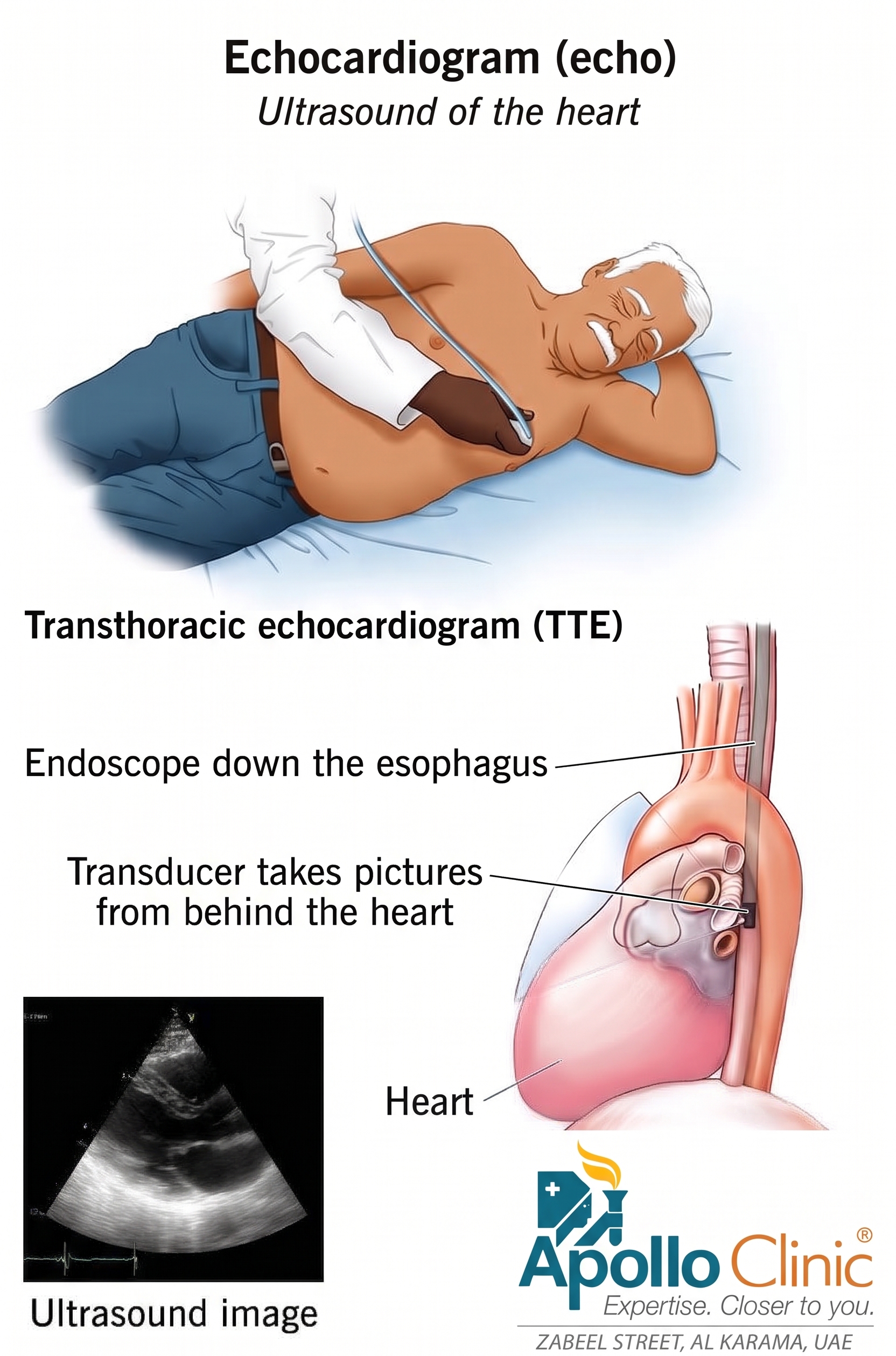
An echocardiogram is a noninvasive heart ultrasound that uses high-frequency sound waves to create moving pictures of the heart as it beats. It can help healthcare providers assess heart structure, pumping ability and blood flow, including blood flow through the heart valves.
The most common type is a transthoracic echocardiogram (TTE), performed by placing a transducer on the chest. Another type, a transesophageal echocardiogram (TEE), obtains images from inside the body and is performed with sedation.
What Is an Echocardiogram Used For?
Cardiologists may order an echocardiogram for several reasons, including to:
Find a possible cause of symptoms.
Confirm or evaluate a diagnosis.
Assess heart structure and pumping function.
Monitor a heart condition such as valve disease.
Help evaluate the heart before surgery.
Assess the outcome of cardiac treatment or surgery.
The reason for the test depends on the patient’s symptoms, medical history and clinical assessment. At Apollo Clinic Dubai, Dr. Shahoo can advise whether an echocardiogram or another cardiac investigation is appropriate based on the individual clinical situation.
Types of Echocardiograms
1. Transthoracic Echocardiogram (TTE)
TTE is the most common type of echocardiogram. A healthcare professional places a small transducer on the surface of the chest to obtain images of the heart. It is generally quick, noninvasive and painless.
2. Transesophageal Echocardiogram (TEE)
TEE produces images of the heart from inside the body. A small transducer is guided down the throat while the patient is sedated. Because the transducer is closer to the heart, TEE can provide higher-quality images of certain structures, including areas that may be difficult to see clearly with a standard TTE.
3. Exercise Stress Echocardiogram
An exercise stress echocardiogram assesses how the heart functions when it is working harder. The patient exercises on a treadmill or stationary bike to increase the heart rate while the heart is evaluated.
4. Dobutamine Stress Echocardiogram
A dobutamine stress echocardiogram also evaluates the heart under stress. Instead of exercise, medication is used to increase the heart rate. This may be useful when a patient cannot exercise adequately on a treadmill or bike.
How Does Echocardiography Work?
An echocardiogram uses ultrasound technology to create images of the heart in motion. A transducer sends sound waves through the chest. The waves reflect from heart tissue and structures, producing echoes that the transducer captures and sends to a computer. Software converts these signals into moving images that the healthcare provider can review and store as part of the medical record.
Different imaging techniques can provide additional information about the heart and blood flow, including:
2D ultrasound: Produces two-dimensional images that can be used to assess the heart’s structures.
3D ultrasound: Creates three-dimensional views and can show cardiac structures from different angles.
Doppler ultrasound: Assesses the speed and direction of blood flow and can help evaluate heart valves and cardiac function.
Strain imaging: Evaluates how the heart muscle contracts and relaxes and may help identify early changes in cardiac function.
Contrast imaging: Uses an injected contrast agent in selected cases to highlight cardiac structures and improve image detail.
Unlike X-rays and CT scans, echocardiography does not use ionizing radiation.
How Should I Prepare for an Echocardiogram?
Preparation depends on the type of echocardiogram. Your healthcare provider will give you specific instructions. Depending on the test, you may be asked to:
Avoid caffeine, alcohol, tobacco or food for a specified period before the test.
Adjust or stop certain medicines only if specifically instructed by your healthcare provider.
Wear comfortable clothing and shoes if you are having an exercise stress echocardiogram.
Arrange for someone to drive you home if you will be sedated.
For many echocardiograms, you may need to undress from the waist up and wear a hospital gown.
What Happens During an Echocardiogram?
A cardiac sonographer usually performs the echocardiogram, with the study interpreted and clinically correlated by the appropriate healthcare professional. A standard echo commonly takes about 40 to 60 minutes, while a TEE may take up to 90 minutes. The exact process depends on the type of test.
You are helped into a comfortable position, usually lying on an examination table for a TTE.
Ultrasound gel may be applied to the chest to help transmit sound waves.
Small electrodes may be placed on the chest to monitor heart rate and rhythm with an EKG.
The transducer is moved over different areas of the chest to obtain images of the heart.
You may be asked to remain still, change position, breathe in certain ways or briefly hold your breath to improve the images.
For a TEE, a numbing solution and sedation are used before the transducer is guided into the esophagus.
After the test, patients who received sedation may need observation before leaving.
Is an Echocardiogram Safe?
Echocardiography is generally considered safe and is usually painless. A standard TTE has no known risks. Other types of echocardiography have specific considerations.
Exercise stress echocardiography can reproduce symptoms such as chest pain or shortness of breath during exertion.
TEE can cause a mild sore throat for a few days.
Contrast agents used in selected echocardiograms can cause an allergic reaction.
Your healthcare provider should explain the relevant risks and answer questions before the procedure.
What Happens After an Echocardiogram?
For most echocardiograms, patients can return home after the test. If sedation was used for a TEE, you will need someone to drive you home, and you should not drive for 24 hours.
The doctor who ordered the echocardiogram will explain when the results are available and may arrange a follow-up appointment or phone consultation. It is best to review the report with your healthcare provider rather than interpreting medical findings on your own.
What Can an Echocardiogram Detect?
An echocardiogram provides moving images that help a healthcare provider evaluate the heart’s structure and function. Depending on the clinical situation, an echocardiogram may help identify or evaluate:
Blood clots in the heart chambers.
Congenital heart disease.
Cardiomyopathy and other conditions affecting the heart muscle.
Certain heart tumors or masses.
Infective endocarditis and abnormalities involving heart valves.
Pericarditis and changes involving the sac around the heart.
Thoracic aortic aneurysm.
Heart valve disease and abnormalities in how valves open or close.
The meaning of an echocardiogram result depends on the full clinical picture. Your cardiologist can explain whether a finding is normal, requires monitoring or needs further evaluation or treatment.
When Should I Contact My Doctor After an Echo?
Contact the doctor who ordered the test if you have questions about preparation, experience a reaction or side effect after the procedure, or have concerns about your results or recommended next steps.
Echocardiogram and Cardiology Care at Apollo Clinic Dubai
At Apollo Clinic Dubai, echocardiography forms part of cardiovascular assessment when clinically indicated. Patients seeking heart evaluation in Dubai can consult Dr. Shahoo, Cardiologist, to discuss symptoms, cardiovascular risk factors, diagnostic needs and appropriate follow-up.
If you have been advised to undergo an echocardiogram, your cardiologist can explain why the test is needed, what type of echocardiogram is appropriate and what the results may mean for your care.
Book a Cardiology Appointment at Apollo Clinic Dubai
To book an appointment at Apollo Clinic Dubai, call +971 4 6056056 or send a WhatsApp message to +971 54289 6804.
Apollo Clinic Dubai provides accessible outpatient healthcare, and patients can discuss their cardiovascular concerns with the appropriate medical team.
Frequently Asked Questions About Echocardiograms
What is an echocardiogram?
An echocardiogram is an ultrasound test that creates moving images of the heart and helps assess its structure, pumping function, valves and blood flow.
Is an echocardiogram painful?
A standard transthoracic echocardiogram is generally painless and noninvasive. Some other types, such as TEE, involve additional preparation and sedation.
How long does an echocardiogram take?
A standard echocardiogram commonly takes about 40 to 60 minutes. A transesophageal echocardiogram may take up to 90 minutes.
Does an echocardiogram use radiation?
No. Echocardiography uses ultrasound sound waves rather than ionizing radiation.
Can I drive after an echocardiogram?
For most routine echocardiograms, patients can drive themselves home. After a TEE involving sedation, someone should drive you home and you should not drive for 24 hours.
Who should I consult for an echocardiogram in Dubai?
A cardiologist can assess your symptoms and medical history and determine whether an echocardiogram is appropriate. At Apollo Clinic Dubai, patients can consult Dr. Shahoo, Cardiologist.
Medical Disclaimer
This article is intended for general health information and does not replace an individual medical consultation, diagnosis or treatment plan. The appropriate type of cardiac test depends on the patient’s symptoms, medical history and clinical assessment.

